What is poor foot alignment?
Poor alignment of the foot can take many forms, with the most commonly being a pronated foot (rolling in at the ankle). This is usually associated with a lowering of the medial longitudinal arch and eversion of the heel/calcaneus (tilting inwards). Another example of poor alignment is the higher arched foot.
The visual appearance of the foot may not be important as how the foot functions during gait. Those with high and low arched feet have been shown to function differently3. Those with lower arched structure tend to have more foot eversion (valgus or pronation) and greater internal rotation of the leg. Those with a higher arch structure were shown to have a poorer ability to absorb shock. In a separate study, the same group of authors4 showed that runners with a higher arch structure had more injuries to the ankle, bone and the injuries were more lateral (outside) whereas the lower arch foot had more injuries to the knee, soft tissues and the injuries were more medial (inside). The maximum amount that a foot pronates (everts) is independent of the height of the arch5 and similar amounts of foot pronation can be seen in those with high and low arch6. This suggests that any sort of foot support or orthoses for the management will need to consider a lot more than supporting the arch or midfoot.
What is now becoming increasingly recognised is that poor alignment can happen in differing amounts in different directions in different feet. As a result of this understanding, no one measure of the way the foot looks or functions is considered appropriate7. There are many different measurements that attempt to quantify the arch, foot alignment or posture or foot shape, but the correlation between each measurement or observation is not good8. A recently developed method is to use a composite score of a number of different observations of different parts of the foot. This ‘Foot Posture Index’9 incorporates observations of foot malignment in different directions (eg height of the arch, amount of heel valgus, amount of medial bulging of the midfoot, the amount of bowing of the achilles tendon, etc). This composite score is considered important to allow for the so-called different “planes of compensation” of the foot. This shows that the poor alignment of the foot can occur in any or all of different body planes (frontal, sagittal and transverse).
Proper alignment of the foot is widely considered as being necessary for normal function during gait.
Poor alignment of the foot is associated with symptoms such as:
- metatarsalgia, arch pain (most commonly plantar fasciitis)
- heel pain (most commonly heel spur syndrome)
- ankle pain, ‘shin splints’ (usually medial tibial stress syndrome)
- overuse syndromes of the knee (usually patello-femoral pain syndrome)
- and a variety of other postural related symptoms.
Foot, leg and back discomfort are common among those with poor foot alignment, especially if they are active or on their feet all day on hard surfaces.
Prefabricated foot orthoses made by Interpod Ltd have been shown to improve the foot malalignment that is associated with these symptoms 1. Not all commonly used prefabricated devices have been shown to change the foot alignment 2.
Poor foot alignment increases risk for tissue damage
Not all those with poor foot alignment (whichever body plane it occurs in) develop problems or symptoms.
A change in foot alignment does not necessarily cause symptoms, but it does increase the risk that it might happen10. Various studies11,12,13 have linked various lower limb alignment characteristics to the risk of developing symptoms. Those runners that pronated more in the rearfoot had more shin splints14 and other lower limb overuse injuries15. Naval recruits had almost twice the risk of getting shin splints if they had a pronated foot type16. Those with bunions and hallux valgus have been shown to have feet that have a lower medial longitudinal arch17,18.
However, not all studies are in agreement that a lower medial longitudinal arch is a sole risk factor for overuse injury19. In a study of 99 grocery store employees, no relationship was found between arch configuration and lower limb pain scores, indicating that the height or configuration of the arch may not necessarily be a factor in lower limb pain20. Other planes of compensation may need further consideration in these types of prospective studies. A lowering of the medial longitudinal arch is compensation in the sagittal plane of the body whereas eversion (rolling in) of the heel is a compensation in the frontal plane. Any attempts to decrease symptoms from foot problems associated with poor foot alignment may need to account for these compensations in both body planes.
Shock absorption
As well as poor alignment, shock absorption also appears to be important in reducing impact loading and improving comfort.
Many studies have shown that shock absorbing insoles can reduce foot pain, when a lack of shock absorption is considered the problem 26-29. Comfort may be an important factor and it is speculated the comfort may delay the onset of fatigue 30.
Foot orthoses decrease the risk by changing foot alignment
Many studies have shown that both prefabricated over-the-counter and custom made foot orthoses or supports can alter the abnormal function of the foot associated with poor foot alignment21,22. Foot orthoses can decrease postural sway23, thereby decreasing the risk for falls.
They been shown to be helpful in selected cases of chronic low back pain24. These types of studies need to be interpreted with caution as foot orthoses are only indicated when foot alignment is a factor in the pain. Landorf and Keenan22 in a comprehensive review of the literature concluded that it is clear that foot orthoses generally have a positive impact on pain and deformity in the feet. They further concluded that foot orthoses do affect position and motion of the foot and lower extremity. While it is still not entirely clear the exact mechanism by which prefabricated foot orthoses exert their effect they are widely used by Podiatrists25.
Current and future research is looking at the “planes of compensation” and how different types of foot orthoses may affect each or all of these. For example, it is highly likely that that those feet that compensate most in the sagittal plane (by collapse of the medial longitudinal arch) will respond most to those foot orthoses that have more support for the midfoot. Another example is that in almost all feet that pronate excessively, as well as the midfoot collapse, the rearfoot everts or goes into valgus. These feet will need wedging under the rearfoot and are not likely to be affected by foot orthoses or supports that merely support the arch or midfoot.
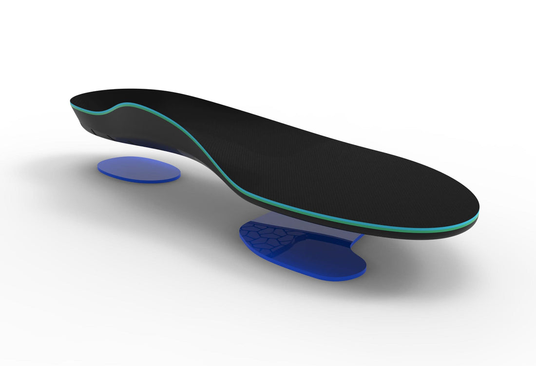
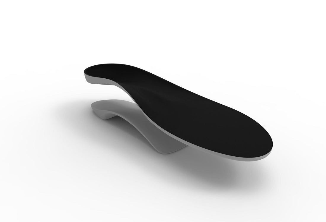
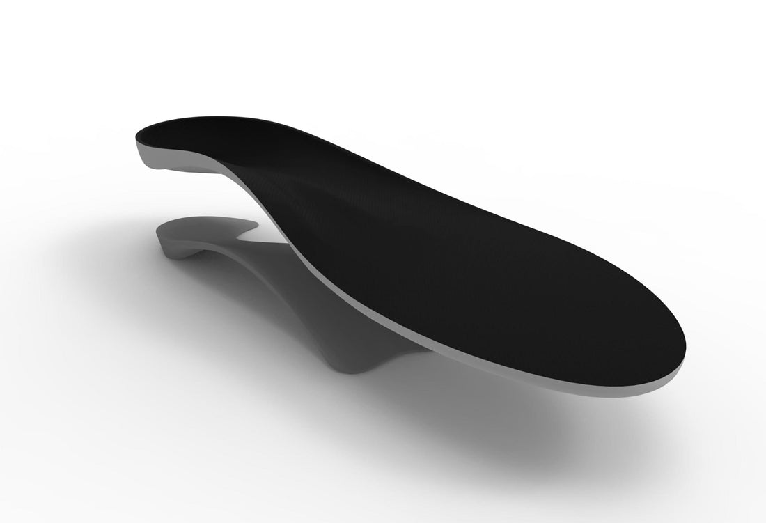
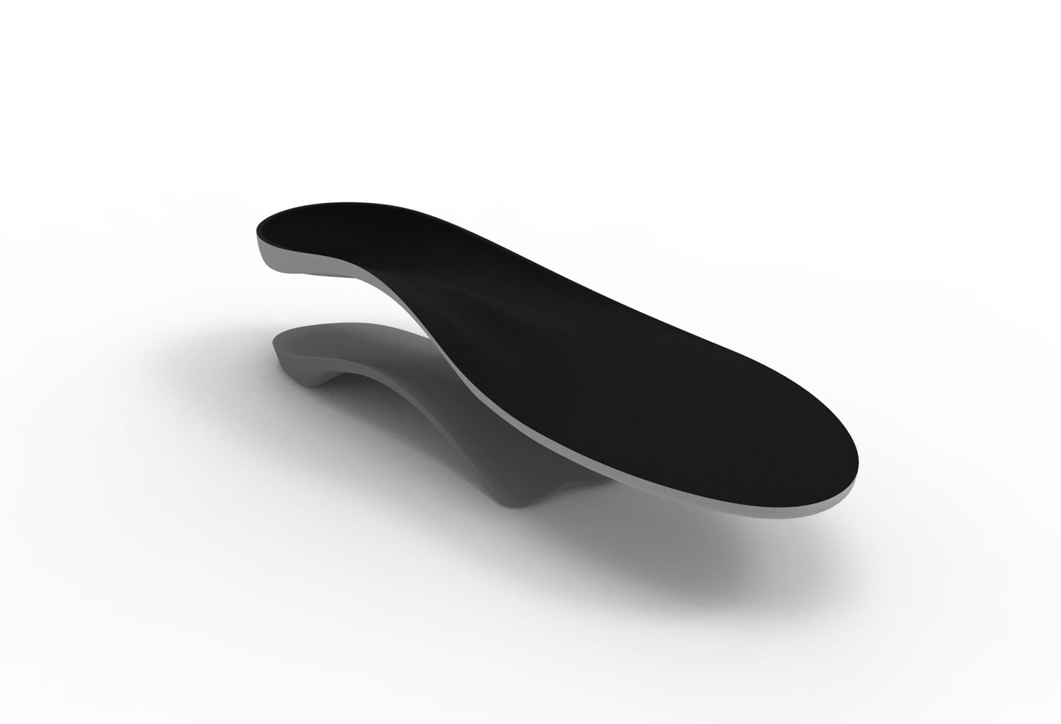
Interpod devices change foot alignment and support the midfoot and absorbs shock
While a lot still remains unclear about how foot orthoses work, they are widely used. They do reduce the incidence of injury to the lower limb, they do change alignment of the foot, they do improve shock absorption and comfort. They also probably have influences by altering sensory feedback. What is also clear is that not all types of foot orthoses can do all of these, all of the time in all people.
As lower arched feet have been shown to have more calcaneal eversion (valgus)3,4, this shows the importance of these feet having the calcaneus position corrected with wedging as well as some midfoot support for the medial longitudinal arch, ie support or control for the different ‘planes of compensation’.
Research at La Trobe University1 has shown that the range of prefabricated foot orthoses by Interpod both increases the height of the medial longitudinal arch and changes the angle of the rearfoot.
References
1. Payne CB, Oates M, Mitchell A: The response of the foot to prefabricated orthoses of different arch height. Australasian Journal of Podiatric Medicine 36(1)7-12 2002
2. Payne CB, Oates M, Noakes H: Static stance response to different types of foot orthoses. Journal of the American Podiatric Medical Association (in press)
3. Williams DS, McClay IS, Hamill J, Buchanan TS: Lower extremity kinematic and kinetic differences in runners with high and low arches. Journal of Applied Biomechanics 17:153-163 2001
4. Williams DS, McClay IS, Hamill J: Arch structure and injury patterns in runners. Clinical Biomechanics 16:341-347 2001
5. Hamil J, Bates BT, Knutzman KM: Relationship between selected static and dynamic lower extremity measures. Clinical Biomechanics 4:217-425 1989
6. Nawoczenski DA, Saltzman CL, Cook TM: The effect of foot structure on the three dimensional kinetic coupling behaviour of the leg and rearfoot. Physical Therapy 78:404-416 1998
7. Razeghi M, Batt ME: Foot type classification – a critical review of current method. Gait and Posture 15:282-291 2002
8. Payne CB & Aquino A: Correlation between different measures of the flat or pronated foot. Journal of Orthopaedic and Sports Physical Therapy 31(5)159 2001
9. Redmond A, Burns J, Crosbie J, Ouvrier R, Peat J: An initial appraisal of the validity of a criterion based, observational clinical rating system for foot posture. Journal of Orthopaedic & Sports Physical Therapy 2001, 31:160
10. Payne CB: Is excessive pronation of the foot really pathologic. Australasian Journal of Podiatric Medicine 33:7-10 1999
11. Busseuil C, Freychat P, Guedj EB, Lacour JR: Rearfoot-forefoot orientation and traumatic risk for runners. Foot and Ankle International. 19:32-37 1998
12. Cowan DN, Jones BH, Frykman PN: Lower limb morphology and risk of overuse injury among male infantry trainees. Medicine and Science in Sports and Exercise 28:945-952 1996
14. Cowan DN, Jones BH, Robinson JR: Foot morphological characteristics and risk of exercise related injury. Archives of Family Medicine 2:773-777 1992
15. Vitasalo JT, Kvist M: Some biomechanical aspects of the foot and ankle in athletes with and without shin splints. 11:125-129 1983
16. Messier SP, Pittala KA: Etiologic factors associated with selected running injuries. Medicine and Science in Sports and Medicine 20:501-505 1988
17. White S: Risk of medial tibial stress syndrome in Naval recruits. Unpublished Honours thesis. La Trobe University, Australia 2001
18. Inman VT: Hallux valgus – a review of aetiological factors. Orthopaedic Clinics of North America 5:863-887 1976
19. Kalen V, Brechner A: Relationship between adolescent bunions and flat feet. Foot & Ankle 8:331-336 1988
20. Ilahi OA, Kohl HW: Lower extremity morphology and alignment and risk of overuse injury. Clinical Journal of Sports Medicine 8:38-42 1998
21. Hogan MT, Staheli LT: Arch height and lower limb pain – an adult civilian study. Foot and Ankle International 23:43-47 2002
22. Bates BT, Osternig LR, Mason B, James LS: Foot orthotic devices to modify selected aspects of lower extremity mechanics. The American Journal of Sports Medicine 7:338-342 1979
23. Landorf KB, Keenan A: Efficacy of foot orthoses – what does the literature tell us? Journal of the American Podiatric Medical Association 90:149-158 2000
24. Oschendorf DT, Mattacola CG, Arnold BL: Effect of orthotics on postural sway after fatigue of the plantar flexors and dorsiflexors. Journal of Athletic Training 35:26-30 2000
25. Dananberg HJ, Guiliano M: Chronic low back pain and its response to custom made foot orthoses. Journal of the American Podiatric Medical Association 89:109-117 1999
26. Landorf K, Keenan AM, Rushworth RL: Foot orthoses prescription habits of Australian and New Zealand Podiatric Physicians. Journal of the American Podiatric Medical Association 4:174-183 2001
27. Tooms RE, Griffin RW, Green P et al: Effect of viscoelsatic insoles on pain. Orthopaedics 10:1143-1148 1987
28. Voloshin A, Wosk J: Influence of artificial shock absorbers in human gait. Clinical Orthopaedics 160:52-57 1981
29. Pratt DJ, Rees PH, Rodgers C: Assessment of some shock absorbing insoles. Prosthetic and Orthotics International 10:43-49 1986
30. Sherman RA, Karstetter KW, May H, Woerman AL: Prevention of lower limb pain in soldiers using shock absorbing orthotic inserts. Journal of the American Podiatric Medical Association 86:117-122 1996
31. Nigg BM, Nurse MA, Stefanyshyn DJ: Shoe inserts and orthotics for sport and physical activity. Medicine and Science in Sports and Exercise 31(suppl):S421-S428 1999
Prepared on behalf of Interpod Ltd by Craig Payne, Department of Podiatry, School of Human Biosciences, La Trobe University, Bundoora, Vic 3086 Australia. Email: c.payne@latrobe.edu.au