Effects of a plantar fascial groove in a foot orthotic on the windlass mechanism
Department of Podiatry, School of Human Biosciences, La Trobe University, Melbourne, Australia
Address for correspondence:
Craig Payne
Department of Podiatry
School of Human Biosciences
La Trobe University
Bundoora, Vic 3083
Australia
Phone: (+61) (3) 9479 5820
Fax: (+61) (3) 9479 5784
Email: c.payne@latrobe.edu.au
Abstract
The windlass mechanism is one way in which the foot can support itself.
This study investigated the force needed to establish the windlass mechanism during static stance in 25 subjects while barefoot and while standing on two different foot orthoses, which were identical except for one having a plantar fascial groove. The force to establish the windlass mechanism was significantly lower (p<0.001) with the foot orthoses with the plantar fascial groove. It is assumed that lowering the force to establish the windlass mechanism will improve gait efficiency.
Introduction
Foot orthoses are widely used to treat biomechanical dysfunction of the lower extremity.
Several theoretical frameworks underpin how foot orthoses alter lower limb kinematics and kinetics with there being no consensus of understanding of the reasons for there clinical effectiveness1,2,3. Foot orthoses may achieve their clinical effect by support the medial longitudinal arch, they may invert the rearfoot and affect skeletal alignment, they may work by altering sensory input or by facilitating the windlass mechanism.
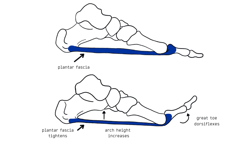
The windlass mechanism, originally described by Hicks4 is one mechanism by which the foot can raise the arch, supinate the rearfoot and become a more stable structure. As the hallux dorsiflexes during gait, the plantar fascia is pulled around the first metatarsal head (the windlass) causing the metatarsal to plantarflex and the medial longitudinal arch to be raised. If it was possible to use foot orthoses to lower the force that is needed to establish the windlass mechanism, it is assumed that this is desirable as it should facilitate the foot being able to support itself. The aim of this project was to compare the force during static stance to establish the windlass mechanism of identically shaped foot orthoses with and without a plantar fascial groove.
Methods
Approval for this study was given by the Faculty of Health Sciences Human Ethics Committee.
Healthy asymptomatic subjects who were free of foot pathology that would interfere with normal first metatarsophalangeal joint dysfunction were recruited for the study.
An apparatus was constructed which consisted of a platform for subjects to stand on in their normal angle and base of gait. The platform was hinged in the area of the first metatarsophalangeal joint so it could be elevated to any desired angle (fig one). A screw mechanism could fix the angle and the force plantarflexing the hallux was measured in Newtons with a Memesion force gauge. The force that was plantar flexing the hallux was considered to be the force that was needed to establish the windlass mechanism. Previous testing showed the device was reliable 5.
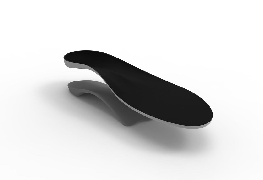
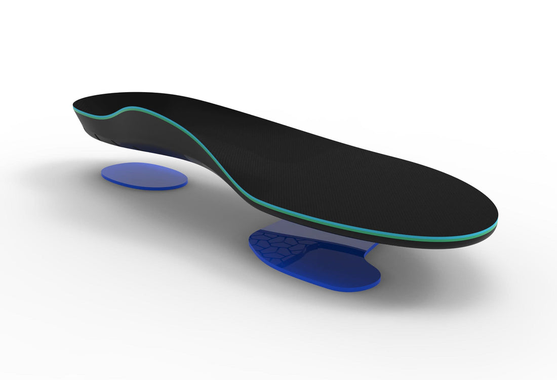
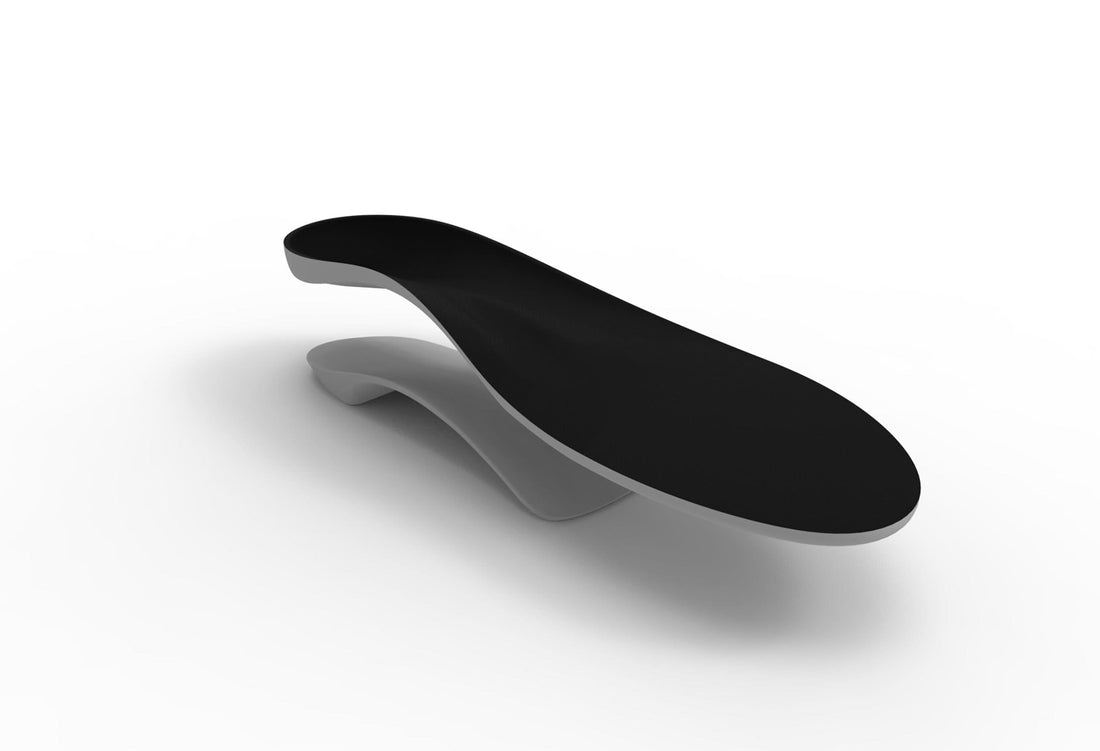
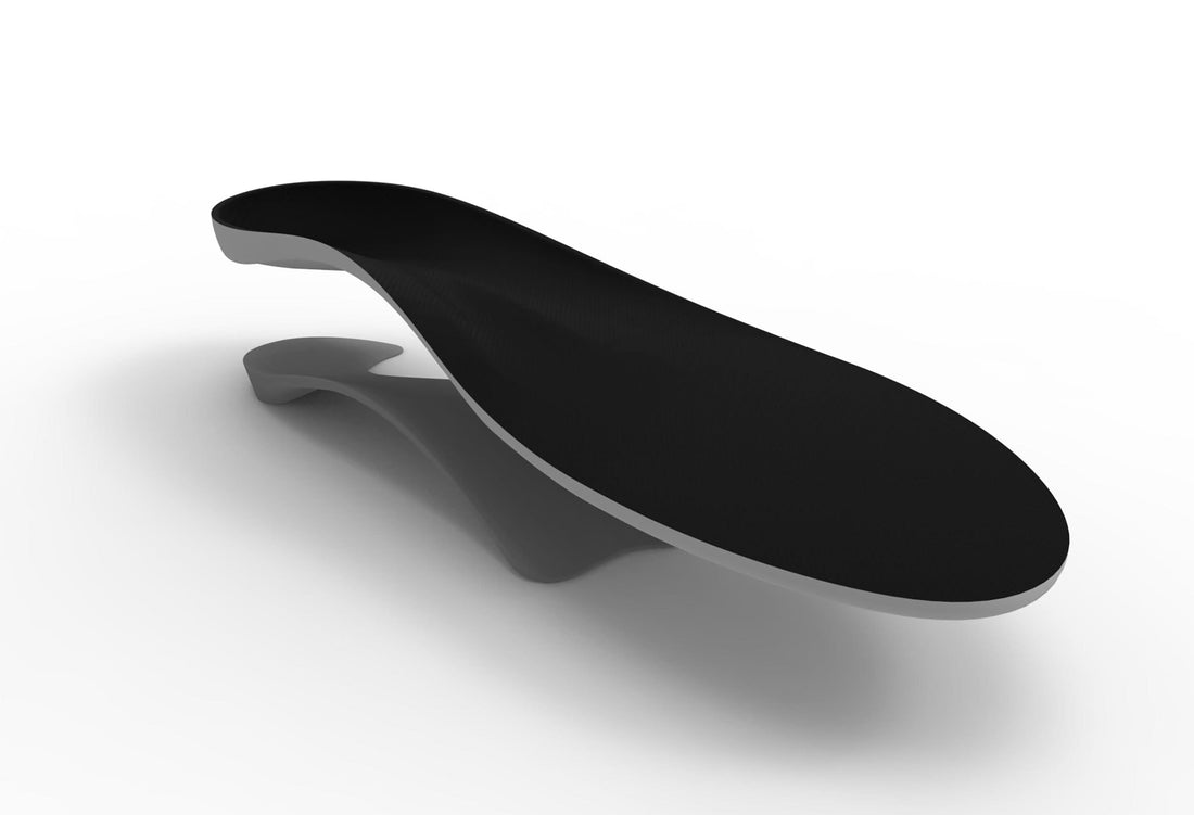
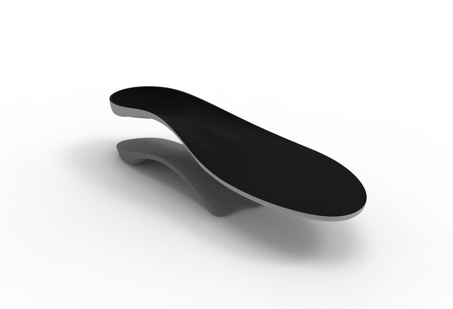
Two prefabricated foot orthoses were used. They were identical with the exception of one having a groove for the plantar fascia. They were constructed of 3mm thick polypropylene (fig two).
The procedure for testing involved subjects standing on the platform in their angle and base of gait, with the long axis of the foot to be tested placed parallel with the platform. The centre of the medial side of the first metatarsophalangeal joint was identified by dorsiflexing the joint several times. The foot was then moved so that it was centred directly over the hinge of the platform. The hinged of the platform was attached to a dial with marks place at different degrees. The hinged platform was elevated to measure the force at 18, x, x degrees of dorsiflexion. The procedure was carried out three times at each angle with the subject barefoot and standing on both the two different foot orthoses. The mean of each of the three tests at each angle was used for the analysis.
Results
The study was performed on the left foot of 25 subjects (mean age: 24.8 years; mean weight: 68.8 kgs; 5 male; 10 female). The mean force (Newtons) under the hallux at each degree of dorsiflexion is reported in table one.
Table One: Force under the hallux (Newtons) at each of the 3 angle of hallux dorsiflexion and 3 different conditions. Using Friedman’s test for a one sample repeated measures design, the results were all significantly different (p<0.001). Post hoc analysis using a paired t-test shows that the different was significant between all three conditions.
Discussion
This study has shown that in healthy subjects that a standard foot orthoses with a groove to accommodate the plantar fascia is effective at lowering the force to establish the windlass mechanism compared to an identically shaped foot orthoses without a groove.
The windlass mechanism is one means by which the foot is able to support itself. The tension that is established in the plantar fascia during hallux dorsiflexion as the heel comes off the ground, raising the medial longitudinal arch and inverting the rearfoot. It could be assumed that if the force that is needed to establish the windlass mechanism is lowered, then this should facilitate gait and stability of the foot. The reduction seen in this study in the force needed to establish the windlass mechanism with a plantar fascial groove could be considered desirable for gait efficiency.
This study needs to be interpreted in the context of limitation of the force to dorsiflex the hallux during gait is assumed to be a proxy for a measure of the force needed to establish the windlass mechanism. The measurement of this force is also done in static stance with the dorsiflexion of the hallux assumed to approximate what happens during dynamic function. Despite these limitations this study has shown that a groove to accommodate the plantar fascia may be effective in improving gait efficiency.
References
1. Payne CB: The past, present, and future of podiatric biomechanics. J Am Podiatr Med Assoc 1998 88: 53-63
2. Lee W: Podiatric biomechanics: an historical appraisal and discussion of the Root model as a clinical system of approach in the present context of theoretical uncertainty. Clinics in Podiatric Medicine & Surgery. 18(4):555-684, 2001
3. Nigg BM, Nurse MA, Stefanyshyn DJ: Shoe inserts and orthotics for sport and physical activities. Medicine and Science in Sports and Exercise. 31(7; Suppl): 421-428 1999
4. Hicks JH: The mechanics of the foot II – The plantar aponeurosis and the arch. Journal of Anatomy 88:25-31 1954
5. Payne CB: Measuring the force to establish the windlass mechanism in the foot (in press)


