The response of the foot to prefabricated orthoses of different arch heights
Craig Payne1, Mathew Oates2 & Alison Mitchel3
(1) Lecturer (2) Associate Lecturer (3) Research Assistant
Department of Podiatry, School of Human Biosciences,
La Trobe University, Melbourne, Australia
Address for correspondence:
Craig Payne
Department of Podiatry
School of Human Biosciences
La Trobe University, Vic 3086, Australia
Phone: (+61) (3) 9479 5820
Fax: (+61) (3) 9479 5784
Email: c.payne@latrobe.edu.au
Word count: 2553 (excl abstract and refs)
Abstract
A number of different design parameters are used in foot orthoses, one of which is the height of the arch.
The aim of this study was to investigate the response of the foot to prefabricated foot orthoses of different arch heights. Sixteen subjects stood in static stance on six different pairs of foot orthoses and changes in navicular height and frontal plane calcaneal angle were measured. All devices resulted in statistically significant changes in the frontal plane calcaneal angle and height of the navicular. The changes to the calcaneal angle were correlated to the amount of force needed to supinate the foot. The changes in navicular height were correlated to the posture of the foot. Further work is needed to determine if these changes are related to dynamic changes and clinical outcomes.
Introduction
Foot orthoses are widely used to treat lower limb biomechanical dysfunction.
A number of decisions have to be made as to which of a multitude of variables need to be considered in the prescription of foot orthoses for this purpose. However, very little evidence is available to guide clinicians in this decision making process. This is further complicated by the kinematic response to foot orthoses being considered as subject specific, in that no two individuals will respond to the same foot orthotic1,2,3. The subject specific variables that determine a kinematic response to foot orthoses have not, as yet, been empirically determined.
Investigating the link between subject specific variables and the different variables that can be incorporated into foot orthoses is complex due to the large numbers of both and the different individual preferences by clinicians. One such variable in the use of foot orthoses is the height of the medial longitudinal arch of the device. It has not been previously reported how subjects respond to the same type of foot orthoses manufactured with different arch heights. The aim of this project was to determine the static stance response of the foot to prefabricated foot orthoses available in different arch heights.
Methods
Subjects were recruited from the undergraduate student population and staff at La Trobe University.
Informed consent was obtained from each participant and the project was approved by the Faculty of Health Science’s Human Ethics Committee. Subjects were recruited who did not have any structural or functional problem of the lower limb that prevented them from weightbearing on different types of foot orthoses.
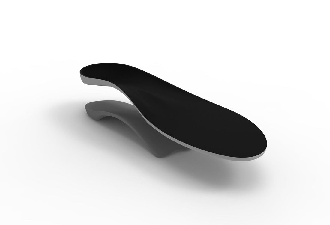
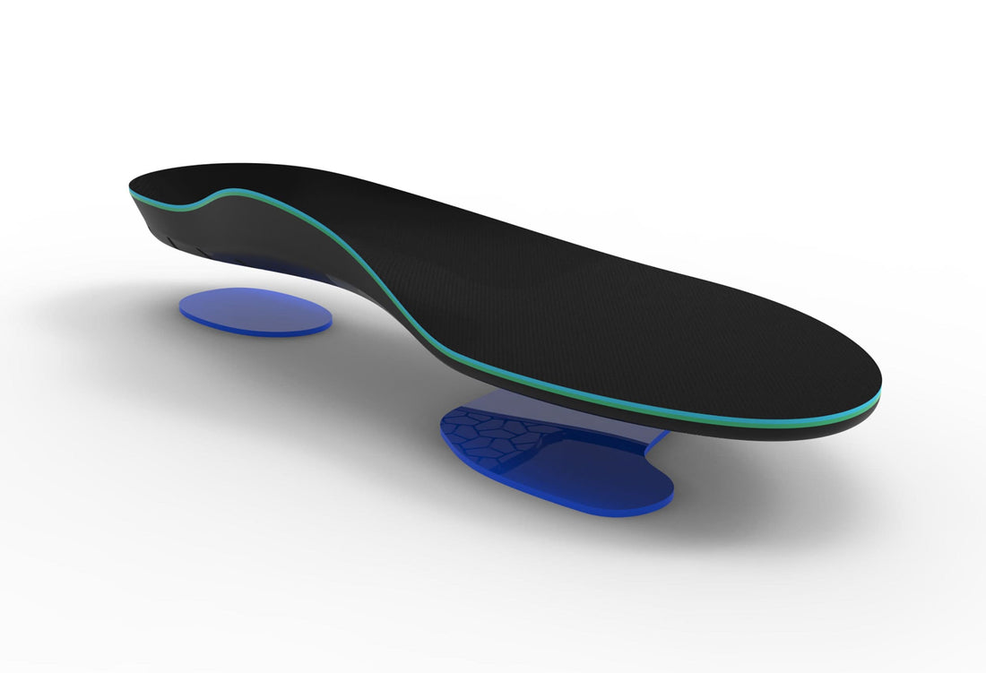
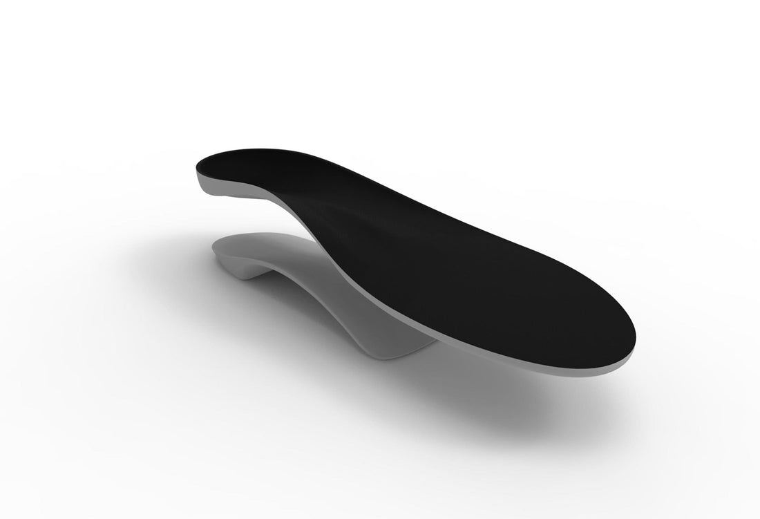
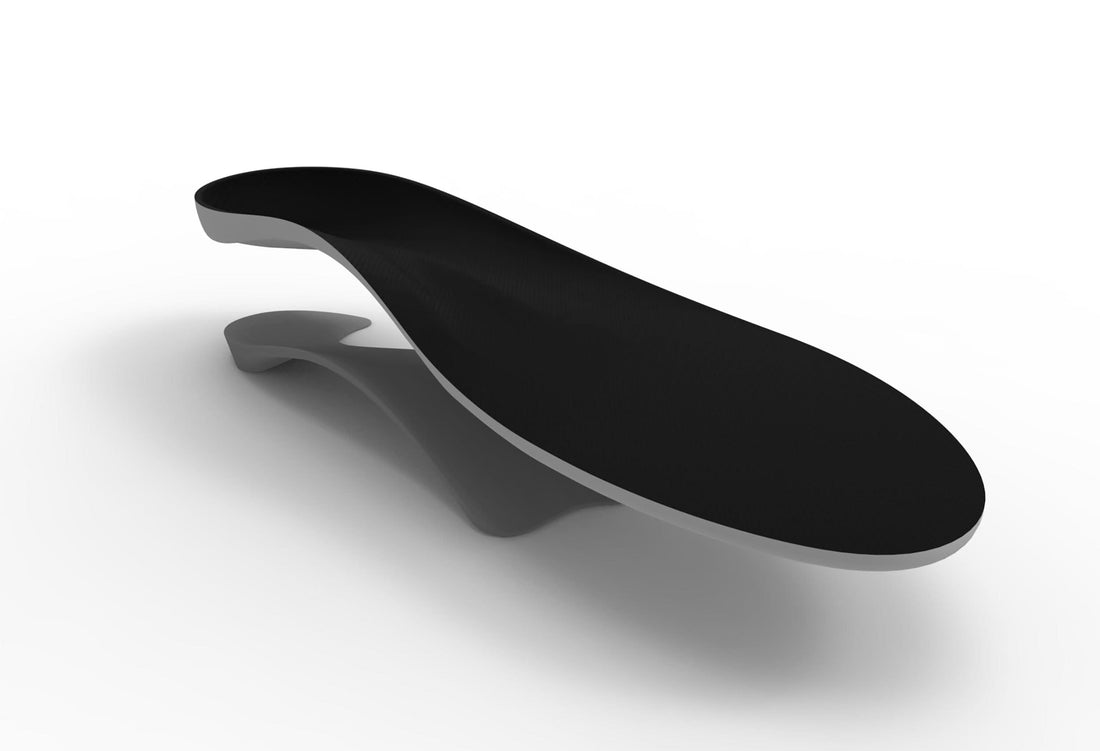
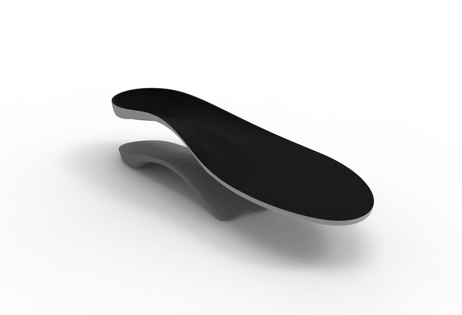
Six prefabricated foot orthoses from the one manufacturer were used so consistency was achieved in the shape of the foot orthoses with the exception of the differing arch heights. Three different plastic (rigid) metatarsal head length devices with rearfoot posts with an identical shape and profile, but with a different arch height (Control Tech Flex 4, 6 and 8 degrees), two soft full-length devices, each with a different arch height (Control Tech Soft 4 and 6 degrees) and a soft metatarsal head length device that is cut down to fit in dress shoes (Slim Tech Soft) (figs one and two).
Canvas shoes were cut down so that the foot orthoses could be placed in the shoes and subjects could be measured standing on the different foot orthoses (fig three). The posterior and medial aspects of the shoes were cut out so that changes in navicular height and frontal plane calcaneal angle could be measured. The resting calcaneal stance angle was measured using a digital inclinometer (Spi-Tronic™) with no orthoses and with each of the orthoses (fig four). The navicular height was measured using digital callipers (Mitutoyo™) with no orthoses and with each of the orthoses (fig five). The skin lines for the measurements were placed by one experienced clinician and changes were independently measured by two experienced clinicians. The mean of the two clinicians was used for the analysis.
To determine foot structure and type, the Foot Posture Index (FPI)4,5,6 was used. As many of the measurements of the foot and lower limb have been shown to have some degree of unreliability associated with them7 the FPI uses 8 observations of the foot and assigns a score of –2 (supinated) to +2 (pronated) to each observation depending on the position of the foot. These weightbearing observations include palpation of the head of the talus for congruence of the talonavicular joint, comparison of the curves above and below the lateral malleoli, bowing of the achilles tendon (Helbing’s sign), frontal plane position of the calcaneus (inverted/everted), bulging in the region of the talonavicular joint, height and congruence of the medial longitudinal arch, congruence of the lateral border of the foot, and abduction/adduction of the forefoot on the rearfoot. A composite score of –1 to +4 is considered to represent a relatively normal foot posture. A score from +5 to +9 is considered mildly pronated and a score above +10 is considered highly pronated (maximum score is +16). A score of –2 to –6 is considered supinated and below –6, highly supinated (lowest score is –16). Two experienced clinicians independently determined the FPI, with the mean of the two being used for analysis.
The composite FPI score was correlated to changes in frontal plane angle and navicular height with the six different devices. In addition 3 of the 8 items that make up the FPI (calcaneal inverted/everted, bulging in the region of the talonavicular joint and congruence of the medial longitudinal arch) were also used to correlate to changes with the foot orthoses. These three were chosen as they are assumed to represent positions of the foot in the frontal plane (calcaneal inversion/eversion), sagittal plane (medial longitudinal arch height) and transverse plane (bulging in talonavicular region).
To determine the amount of force needed to supinate the rearfoot, a previously described mechanical supination resistance device8,9,10 was used (fig six). This device consisted of a non-stretchable woven fabric that was fixed to the base of a platform lateral to the foot in the region of the calcaneocuboid joint. It passed under the foot 5 medial to the talonavicular joint, then proximally to be attached to a pulley system and a Mecmesin® force gauge that measured the force in Newtons. The pulley system was used to apply a force to just overcome the inertia needed to invert the calcaneus, as the investigator observed a bisection that was placed on the posterior aspect of the calcaneus. Ten trials were collected from each foot of the subjects with the mean used for the analysis. The device has been previously reported to be reliable9.
Repeated measures ANOVA was used to determine the differences in frontal plane calcaneal angle and navicular height from the barefoot condition and the use of the six difference devices. Bonferroni post hoc testing was used to determine where the difference occurred. Pearson’s correlation coefficient was used to determine the association of the FPI and supination resistance to the changes in the frontal plane position of the calcaneus and the height of the navicular for each of the orthoses. Intraclass correlation coefficients (type 2,1) were used to determine the intra-rater reliability of the two clinicians in determining the FPI and measuring the changes in the frontal plane calcaneal angle and the height of the navicular.
Results
Many studies have shown that both prefabricated over-the-counter and custom made foot orthoses or supports can alter the abnormal function of the foot associated with poor foot alignment(21),(22). Foot orthoses can decrease postural sway(23), thereby decreasing the risk for falls.
They been shown to be helpful in selected cases of chronic low back pain(24). These types of studies need to be interpreted with caution as foot orthoses are only indicated when foot alignment is a factor in the pain. Landorf and Keenan(22) in a comprehensive review of the literature concluded that it is clear that foot orthoses generally have a positive impact on pain and deformity in the feet. They further concluded that foot orthoses do affect position and motion of the foot and lower extremity. While it is still not entirely clear the exact mechanism by which prefabricated foot orthoses exert their effect they are widely used by Podiatrists(25).
Current and future research is looking at the “planes of compensation” and how different types of foot orthoses may affect each or all of these. For example, it is highly likely that that those feet that compensate most in the sagittal plane (by collapse of the medial longitudinal arch) will respond most to those foot orthoses that have more support for the midfoot. Another example is that in almost all feet that pronate excessively, as well as the midfoot collapse, the rearfoot everts or goes into valgus. These feet will need wedging under the rearfoot and are not likely to be affected by foot orthoses or supports that merely support the arch or midfoot.
Discussion
This study investigated the static stance change in the foot in response to the use of prefabricated foot orthoses with different arch heights from one manufacturer (Interpod Ltd, Melbourne, Australia).
The use of these measurements are based on the assumption that realignment of the foot is the aim for the use of foot orthoses that are designed to alter foot function, which recent work suggests may not necessarily be the only aim of foot orthoses1,2. Any study of the type presented here needs measurements that are reliable. The measurements that were used in this study have previously been shown to be generally unreliable7,11,12,13. In our study the mark that was placed on the skin to represent navicular height and calcaneal angle was not removed for subsequent measurements when changes with the foot orthoses where measured, which would have improved reliability. The reliability of the three measurements used in this study (FPI, calcaneal angle and navicular height) had ICC’s of 0.83 to 0.96, indicating that the measurements are reliable enough for the data to be used for subsequent analysis.
The results have shown that all of the devices within the Interpod range (Control Tech Flex, Control Tech Soft and Slim Tech Soft) resulted in a statistically significant change in the position of the calcaneus in the direction of inversion, as well as a 8 significant increase in the height of the navicular. If the therapeutic aim is to change the calcaneal angle or improve arch height, these prefabricated foot orthoses can achieve this in static bipedal stance. The results presented here are comparable to the prefabricated foot orthoses reported in a previous study that resulted in statistically significant change in the frontal plane angle of the calcaneus in the direction of inversion3. In that study, three prefabricated foot orthoses resulted in a change in the frontal plane angle of the calcaneus and three other prefabricated foot orthoses did not. The design parameter that determined the ability of the orthoses to invert the foot was the use of medial heel. This medial heel wedging would increase the supination moment on the medial side of the subtalar joint axis14. The orthoses used in the study presented here have design features that do the same, which would account for the change in the calcaneal angle in the direction of inversion.
Supination resistance testing has been developed as a method to measure the force needed to supinate the rearfoot in static stance8,9,10. This study has demonstrated a moderate negative correlation between the force needed to supinate the foot (supination resistance) and the change in the frontal plane calcaneal angle for all of the six orthoses (one was not statistically significant). This means that the greater the supination resistance, the less the change in frontal plane angle of the calcaneus. As a manual version of supination resistance testing has been shown to be reliable when used by those experienced in its use10, this finding could possibly be extrapolated to be used clinically to assist in deciding how much change is likely to happen in the static stance change to the frontal plane angle of the calcaneus with foot orthoses. There was also a moderate negative correlation between supination resistance and navicular height changes for the soft orthoses (Control Tech Soft and Slim Tech Soft). 9
This can be interpreted as, for example, when the supination resistance was higher, there was less change in navicular height when using a softer orthoses. For the rigid orthoses (Control Tech Flex) this did not happen, which indicates that changes in navicular height with the use of rigid orthoses was not related to the force needed to supinate the foot.
orthoses. A mild positive correlation was noted for the two rigid devices with the higher arch heights, but this did not quite reach significance. For all the orthoses, bulging in the talonavicular region (as a component of the FPI) was moderately and positively correlated to changes in the navicular height; ie the greater the medial bulging, the greater the change in navicular height.
The results of this study have to be interpreted in the context of a number of limitations. All the subjects were healthy asymptomatic young adults and may not be representative of a pathological population, however the range of FPI values (-1, ‘normal’ to 11.58, severely pronated) and supination resistance values (78.72 to 10 358.47) indicate a wide range of foot types used in the study. All measurements used in this study were done in static bipedal (double limb) stance and not dynamically, so they may not necessarily represent what occurs during dynamic stance. Also, the different outcomes noted between the hard and soft orthoses may have been due to other variables, such as the softer devices have higher flanges and are wider.
Conclusion
In conclusion, this study has shown that the Interpod range of prefabricated foot orthoses can result in a statistically significant change during static stance in the frontal plane position of the calcaneus in the direction of inversion and an increase in the arch height.
We have also shown that the greater the force needed to supinate the foot, the less will be the change in the frontal plane calcaneal angle. Using the Foot Posture Index as a measure of how pronated the foot is, it was also shown that the posture of the foot was not related to how much the calcaneal angle can change, but it is related to how much the height of the navicular changes. Further work is needed to investigate if these static changes are related to dynamic changes and if they are related to clinical outcomes.
References
1. Nigg BM. The role of impact forces and foot pronation: a new paradigm. Clinical Journal of Sport Medicine. 2001, 11:2-9
2 Payne CB & Chuter V: The clash between the science and the theory on the kinematic effectiveness of foot orthoses. Clinics in Podiatric Medicine and Surgery 2001, 18:705-713
3. Payne CB, Oates M & Noakes H: Static stance response to different type of foot orthoses. Journal of the American Podiatric Medical Association (in press)
4. Redmond A, Burns J, Crosbie J, Ouvrier R, Peat J: An initial appraisal of the validity of a criterion based, observational clinical rating system for foot posture. Journal of Orthopaedic & Sports Physical Therapy 2001, 31:160
5. Redmond A, Crosbie J, Peat J, Burns J, Ouvrier R: A new criterion based, composite clinical rating system for the quantification of foot posture – its validation and use in clinical trials. Book of Abstracts, 19th Australasian Podiatry Conference 16-17 May 2001
6. Redmond AC, Burns J, Crosbie J, Peat JK, Ouvrier RA: A rating system for the quantification of foot posture: the Foot Posture Index. (submitted)
7. Menz HB. Clinical hindfoot measurement: a critical review of the literature. The Foot. 1995, 5:57-64
8. Payne CB, Munteanu S, Miller K: Position of the subtalar joint axis and resistance of the rearfoot to supination. Journal of the American Podiatric Medical Association (in press)
9. Payne CB, Noakes H, Oates M, Munteanu S: Resistance of the foot to supination. (submitted)
10. Noakes H & Payne CB: The reliability of the manual supination resistance test (submitted)
11. Vinicombe A, Raspovic A, Menz H: Reliability of Navicular Displacement Measurement as a Clinical Indicator of Foot Posture. Journal of the American Podiatric Medical Association 2001 91: 262-268.
12. Payne CB & Richardson MR: Changes in the measurement of RCSP and NCSP with experience. The Foot 2000; 10:81-84
13. Williams DS, McClay IS: Measurements used to characterise the foot and the medial longitudinal arch – reliability and validity. Physical Therapy 200; 80864-871
12. Payne CB & Richardson MR: Changes in the measurement of RCSP and NCSP with experience. The Foot 2000; 10:81-84
14. Kirby KA: Subtalar joint axis location and rotational equilibrium theory of foot function. Journal of the American Podiatric Medical Association. 2001, 91:465-472.
Table One
Changes in the static stance frontal plane calcaneal angle from no orthoses condition and the different orthoses.

Table Two
Static stance height in the no orthoses condition and the different orthoses.

Table Three
Correlation between supination resistance and Foot Posture Index and changes in the calcaneal angle and navicular height for each of the six prefabricated devices.

Table Four
Correlation between arch height, calcaneal position and midfoot bulging and changes in frontal plane calcaneal angle and navicular height. Arch height, calcaneal position and midfoot bulging are components of the FPI and have a range of –2 (supinated) to +2 (pronated).

Figure 1
The three rigid pre-fabricated foot orthoses with different arch heights used in the study (Control Tech Flex, Interpod Ltd).

Figure 2
The two different types of soft prefabricated foot orthoses used in the study (Control Tech Soft and Slim Tech Soft, Interpod Ltd).

Figure 3
The cut down canvas shoes used in the study so that changes in frontal plane calcaneal angle and navicular height could be measured.

Figure 4
The use of the digital inclinometer to measures changes in the frontal plane calcaneal position with the use of the different foot orthoses

Figure 5
The use of the digital callipers to measure changes in the height of the navicular with the use of the different foot orthoses.

Figure 6
The supination resistance device used to measure the amount of force needed to supinate the rearfoot.


